
Derm Spotlight: Treatment Options for Calcium Deposits in Dog Skin

What is Calcinosis Cutis?
This is an uncommon physiological process characterized by inappropriate mineral deposition into the epidermis (superficial skin), dermis (deep skin), and subcutis (fatty layer underneath the skin) with calcium salts. Calcinosis cutis in dogs is a rare disease usually occurring in large breeds (for example, German Shepherds) and can affect puppies and senior dogs. The condition has also been reported in cats and humans.
What causes Calcinosis Cutis?
The specific cause of the condition is unclear, although there may be a genetic component. The most common cause of this condition is long term steroid administration. It has also been associated in dogs diagnosed with Cushing’s disease. Few cases have been reported in dogs after receiving the medication Amphotericin B.
It has also been associated with cutaneous (skin) exposure to bone meal, landscaping products, barn dusts, and/or ice melters. Rarely, hypoparathyroid dogs and cats that receive intravenous or subcutaneous calcium chloride or calcium gluconate injections have developed calcinosis cutis.
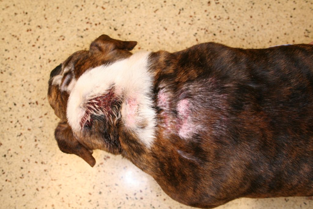
The back of an Old English Bulldog after long term steroids use. You can see hair loss, pink circular raised plaques on the back of the neck and shoulder region.
What does Calcinosis Cutis look like?
Lesions consist of firm, gritty yellowish-tan dark red papules (small bumps) and plaques (large, raised circular). Ulcerated and crusting noted with a superficial gritty mineral material can be seen on the surface. This change may be focal (one spot) to widespread (many areas affected). Areas affected include the back of the neck, back, abdomen, axillae (arm pits), groin. It is often confused with a superficial bacterial infection on the skin.
Symptoms of calcinosis cutis may include
- Skin lesions
- Body Muscle Deficiency
- Blackheads
- Hard lumps under skin, mouth, and footpads
- Loss in weight
- Loss of hair
Calcinosis cutis lesions can become ulcerated and infected, especially if dogs are able to access the area with their mouth or paws.
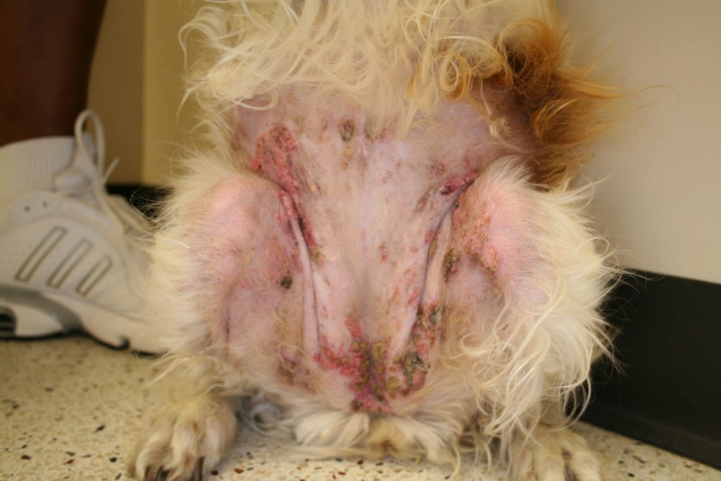
This is the abdomen of a Brittany Spaniel that was diagnosed with Cushing’s disease. You can see the red raised plaques with a yellow-tan gritty surface.
How is Calcinosis Cutis diagnosed?
The condition can be diagnosed by examination and confirmed via biopsy. The biopsy site should be an area of skin without excoriations (scratches) and not ulcerated.
How is Calcinosis Cutis treated?
To treat calcinosis cutis in dogs, veterinary dermatology specialists will want to address underlying conditions first. Successful treatment of underlying conditions often helps to alleviate the skin nodules caused by calcium deposits.
Other treatments may include:
- Medicated shampoos
- Hydrotherapy
- Antibiotics
- Surgery
- DMSO (dimethyl sulfoxide)
If long term steroids are given, these are usually weaned off and discontinued. If steroids are not given, veterinary dermatology specialists typically will ask pet owners about the pet’s environmental exposure to bone meal, landscaping products, barn dusts, and/or ice melters. If an exposure is identified, it should be removed from the pet’s environment.
If Cushing’s disease is suspected, diagnostics will be needed to make this diagnosis. If Cushing’s Disease has already been diagnosed, then better control of this disease is needed to help resolve the calcinosis cutis. After elimination of the cause, topical DMSO can be added to hasten the resolution.
What is the prognosis of Calcinosis Cutis?
It’s important not to let the dog chew, strike, scrape or rub the infectious places on their body. Once the underlying cause has been identified and addressed, calcinosis cutis can be managed and cured with medication. Other treatments can involve non-steroidal medicines to relieve itchiness. When the root cause has been diagnosed, the prognosis is excellent.
If you think your pet may have allergies and need further evaluation by one of our dermatologists please give us a call!
References:
- Campbell, KL, Griffin, CE, Miller, WH. Muller & Kirk’s Small Animal Dermatology, 7th edition. Elsevier St. Louis Missouri 2013.
- Rhodes, KH, Werner, AH. Blackwell’s Five-Minute Veterinary Consult Clinical Companion Small Animal Dermatology, 3rd Edition. John Wiley and Sons 2018.
- Gross, Ihrke,Walder, Affolter. Skin Diseases of the Dog and Cat, Clinical and Histophatologic Diagnosis, 2nd Edition. Blackwell Publishing company 2005.
